
Endometriosis vs. Adenomyosis: Understanding Two Distinct Conditions

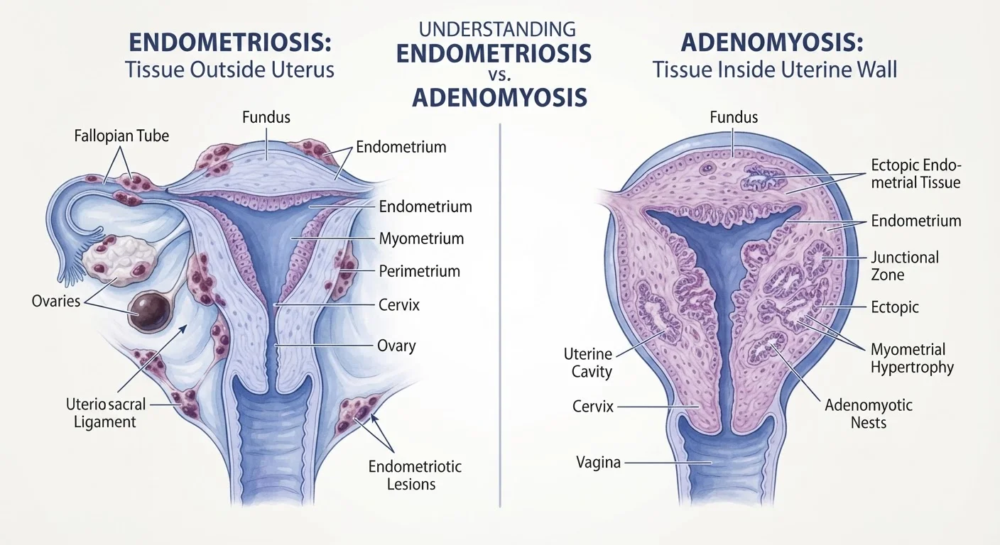
Photo: FeaturingHealth.net | Healthy Living Simplified editorial
Key Takeaways
- Endometriosis involves tissue growing outside the uterus; adenomyosis involves tissue growing into the uterine muscle itself.
- Both conditions can cause significant pelvic pain and menstrual irregularities, making clinical distinction essential.
- Endometriosis is definitively diagnosed via laparoscopy; adenomyosis is typically identified through imaging.
- The two conditions can co-exist in the same person, complicating diagnosis and symptom management.
- Neither condition is a personal failing — both are recognized medical diagnoses requiring professional evaluation.
What Sets These Two Conditions Apart
Endometriosis and adenomyosis are both defined by abnormal growth of endometrial-like tissue, yet they are fundamentally different in where that tissue travels and what damage it causes. Understanding this distinction is clinically meaningful — and it matters for how each condition is diagnosed and managed.
In endometriosis, tissue resembling the uterine lining (the endometrium) implants and grows outside the uterus — commonly on the ovaries, fallopian tubes, the lining of the pelvis, and occasionally on the bladder or bowel. These implants respond to hormonal cycles, swelling and shedding without an exit route, which triggers inflammation, scarring, and adhesions over time.
In adenomyosis, endometrial tissue invades the myometrium — the muscular wall of the uterus itself. Rather than spreading to distant sites, it infiltrates locally, causing the uterine wall to thicken. The result is often a noticeably enlarged, tender uterus and disrupted menstrual flow. For more on how related hormonal conditions compare, see our guide on PCOS and endometriosis.
| Criterion | Endometriosis | Adenomyosis |
|---|---|---|
| Tissue location | Outside the uterus (pelvis, organs) | Inside the uterine muscle wall |
| Primary symptom | Pelvic pain, painful periods | Heavy/prolonged bleeding, uterine pressure |
| Fertility impact | Frequently significant | Can occur; generally less pronounced |
| Uterine size | Usually normal | Often enlarged or tender |
| Primary diagnosis method | Laparoscopy (surgical) | Ultrasound or MRI (imaging) |
| Can co-exist with the other? | Yes — frequently | Yes — frequently |
Symptoms: Overlapping but Distinct
Both conditions produce pelvic pain and menstrual disruption, which is a primary reason they are so frequently confused. However, the character and focus of symptoms differ in ways that can guide clinical suspicion.
Endometriosis is strongly associated with dysmenorrhea (painful periods), deep dyspareunia (pain during or after intercourse), pain with bowel movements or urination during menstruation, and — crucially — subfertility or infertility. Pain often begins before menstruation and continues after it ends. Lesions on the ovaries can form cysts called endometriomas.
Adenomyosis tends to produce heavy or prolonged menstrual bleeding (menorrhagia), a dull pelvic pressure or feeling of uterine heaviness, and cramping that may worsen with age. Fertility impacts are recognized but generally less pronounced than with endometriosis, though significant adenomyosis can affect implantation.
Co-Occurrence Is Common
Symptom overlap is substantial, and both conditions can present mildly or severely regardless of the extent of tissue involvement. A healthcare provider's assessment is the only reliable way to distinguish them.
Diagnosis and Management Approaches
The diagnostic paths for these two conditions diverge significantly, which underscores why accurate identification matters.
Endometriosis has historically required surgical confirmation: a laparoscopy (minimally invasive camera surgery) allows a clinician to directly visualize and biopsy implants. While imaging such as transvaginal ultrasound or MRI can detect endometriomas or deep infiltrating disease, negative imaging does not rule out endometriosis.
Adenomyosis, by contrast, can often be identified through non-invasive imaging. Transvaginal ultrasound and pelvic MRI can reveal characteristic changes in uterine wall thickness and texture. Definitive histologic diagnosis has traditionally required hysterectomy, but imaging has become increasingly reliable for clinical diagnosis.
~10%
Estimated prevalence of endometriosis
The World Health Organization estimates endometriosis affects approximately 10% of reproductive-age women and girls globally.
Up to 7 years
Average diagnostic delay for endometriosis
Multiple studies have reported an average delay of five to seven years between symptom onset and an endometriosis diagnosis, highlighting barriers to timely care.
20–35%
Estimated adenomyosis prevalence in symptomatic women
Prevalence estimates for adenomyosis vary widely in the literature due to historically requiring surgical tissue confirmation, but figures commonly cited range from 20–35% in symptomatic populations.
Management for both conditions focuses on symptom relief and, where relevant, preserving fertility. Hormonal therapies — such as combined hormonal contraceptives, progestins, or GnRH agonists — are commonly used. Surgical options range from excision of endometriotic lesions to, in some cases of adenomyosis, hysterectomy. Treatment plans should always be individualized in consultation with a gynecologist or reproductive endocrinologist.
This article is for general informational purposes only and does not constitute medical advice. Always consult a qualified healthcare professional for diagnosis, treatment decisions, or concerns about your reproductive health.
The content on this site is for informational purposes only and is not a substitute for professional advice. Always consult a qualified professional for guidance specific to your situation.